The Fundamentals of Automatic
X-ray Inspection
for Titanium Applications
Titanium manufacturers have long recognized the need to insure the purity of the titanium chips used in their forging process. As the titanium castings undergo various machining and drilling processes using tungsten tooling, tungsten particles can be introduced into the titanium chips cleaned from the castings. If the tungsten particles are not removed before the titanium chips are used in the next forging process, they will cause stress points in the titanium casting. It is, therefore, critical to remove the tungsten particles before the forging process.
In the early 1980’s, Automatic Defect Recognition (ADR) techniques were introduced to the titanium industry to accomplish the task of removing the tungsten particles. ADR integrates X-ray inspection with machine vision processes. X-rays penetrate the chips, an imaging system generates images for evaluation, and machine vision processes automate analysis of the image and the decision making process.
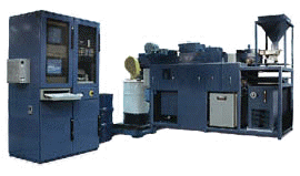 The basic material flow of early titanium X-ray
inspection systems is much the same as today’s systems. Material is loaded into
a hopper. The hopper vibrates to keep the chips from packing together.
Proximity switches at the top and center of the hopper keep a constant level of chips in
the hopper. Chips are spread onto a constantly moving belt which moves the chips
through an X-ray inspection zone. When an inclusion is detected by the ADR process,
the PC gives a signal to stop the belt and vacuum the section of belt which includes the
inclusion. That portion of the belt is then re-inspected, and if all is okay,
inspection continues. Good chips are deposited into a drum or exit conveyor at the
end of the system. (See figure 1.)
The basic material flow of early titanium X-ray
inspection systems is much the same as today’s systems. Material is loaded into
a hopper. The hopper vibrates to keep the chips from packing together.
Proximity switches at the top and center of the hopper keep a constant level of chips in
the hopper. Chips are spread onto a constantly moving belt which moves the chips
through an X-ray inspection zone. When an inclusion is detected by the ADR process,
the PC gives a signal to stop the belt and vacuum the section of belt which includes the
inclusion. That portion of the belt is then re-inspected, and if all is okay,
inspection continues. Good chips are deposited into a drum or exit conveyor at the
end of the system. (See figure 1.)
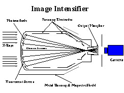 Early titanium X-ray inspection systems used an image intensifier (figure 2),
newvicon camera, particle detection boards, and special circuitry for video enhancement of
the contrast between the titanium chips and the high-density inclusions. The X-ray
tube generated X-rays which were partially absorbed by the chip layer. The X-rays
that pass through the chips strike the fluorescent screen of the image intensifier which
glows in response to the X-rays. The light is magnified and focused on the output
phosphor of the image intensifier. The light is received by the newvicon camera and
scanned. Video information from the camera is sent to the image enhancement boards
for signal processing and the pass/fail decision.0
Early titanium X-ray inspection systems used an image intensifier (figure 2),
newvicon camera, particle detection boards, and special circuitry for video enhancement of
the contrast between the titanium chips and the high-density inclusions. The X-ray
tube generated X-rays which were partially absorbed by the chip layer. The X-rays
that pass through the chips strike the fluorescent screen of the image intensifier which
glows in response to the X-rays. The light is magnified and focused on the output
phosphor of the image intensifier. The light is received by the newvicon camera and
scanned. Video information from the camera is sent to the image enhancement boards
for signal processing and the pass/fail decision.0
While these systems were in fact automatic defect recognition systems, they were able to X-ray and analyze only one section of the belt at a time which slowed the processing speed. They were capable of finding a 0.15" discontinuity; however, blemishes inherent in image intensifiers were occasionally mistaken for contaminants in the titanium chips. These early ADR systems were also limited by other characteristics of image intensifiers such as their ability to generate only 255 grayshades, their reduced image contrast due to X-ray scatter, and their increased potential for geometric distortion.
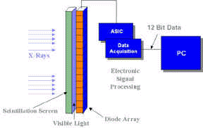 In the late 1980s, linear diode array (LDA) imaging
systems were introduced into the NDT market. ( See figure 3.) The LDA offered more
favorable characteristics in many areas. There are no blemishes inherent in the
system, it is able to generate 4095 grayshades, X-ray scatter has negligible effect on its
image contrast, and geometric distortion is eliminated. While the first LDA systems
were very hardware specific and suffered from decreased reliability and increased
installation and routine maintenance time, today’s systems are technologically
advanced. These systems consist of a standard IBM-architecture PC with Microsoft
Windows NT operating system, an SVGA monitor, and a 1,024-element diode array. They
are highly reliable and easy to upgrade and maintain. With Windows NT and point and
click graphical interface, the operator interface is user friendly.
In the late 1980s, linear diode array (LDA) imaging
systems were introduced into the NDT market. ( See figure 3.) The LDA offered more
favorable characteristics in many areas. There are no blemishes inherent in the
system, it is able to generate 4095 grayshades, X-ray scatter has negligible effect on its
image contrast, and geometric distortion is eliminated. While the first LDA systems
were very hardware specific and suffered from decreased reliability and increased
installation and routine maintenance time, today’s systems are technologically
advanced. These systems consist of a standard IBM-architecture PC with Microsoft
Windows NT operating system, an SVGA monitor, and a 1,024-element diode array. They
are highly reliable and easy to upgrade and maintain. With Windows NT and point and
click graphical interface, the operator interface is user friendly.
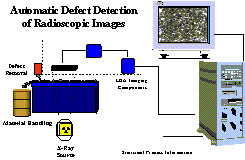 Today’s
titanium X-ray inspection systems use a solid state, high resolution LDA directly coupled
to a cadmium tungstate crystal. This is a greatly simplified image generating
system with excellent image characteristics. The system is operated by a PLC with an
additional PC running Wonderware for the man-machine interface. (See figure
4.) Image analysis is performed by proprietary feature extraction software.
These new processing capabilities provide a major improvement in the ability to
discriminate between real contaminants and larger chips.
Today’s
titanium X-ray inspection systems use a solid state, high resolution LDA directly coupled
to a cadmium tungstate crystal. This is a greatly simplified image generating
system with excellent image characteristics. The system is operated by a PLC with an
additional PC running Wonderware for the man-machine interface. (See figure
4.) Image analysis is performed by proprietary feature extraction software.
These new processing capabilities provide a major improvement in the ability to
discriminate between real contaminants and larger chips.
 As chips are transported through the X-ray inspection zone, the LDA signal is
compared to a pre-determined threshold. (See figure 5.) When the threshold is
exceeded, the software determines whether the detection was caused by a legitimate
contaminant or a larger chip. If the software determines there is a legitimate
high-density inclusion, it is vacuumed off the belt automatically. The vacuumed area
is re-inspected and belt motion resumes. If the software determines it is a large
chip rather than an inclusion, the vacuum sequence is not initiated and the belt motion is
not affected.1
As chips are transported through the X-ray inspection zone, the LDA signal is
compared to a pre-determined threshold. (See figure 5.) When the threshold is
exceeded, the software determines whether the detection was caused by a legitimate
contaminant or a larger chip. If the software determines there is a legitimate
high-density inclusion, it is vacuumed off the belt automatically. The vacuumed area
is re-inspected and belt motion resumes. If the software determines it is a large
chip rather than an inclusion, the vacuum sequence is not initiated and the belt motion is
not affected.1
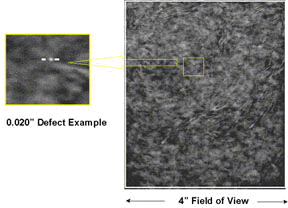
Today’s titanium X-ray inspection systems are capable of finding discontinuities of 0.010" and smaller. The size of the defect to be detected will dictate how magnification, kv, ma, bed depth, belt width or feed rates will be affected. The smaller the defect to be detected, the higher the magnification required, the thinner the bed depth, or the slower the belt speed required. If alloys other than titanium are run, adjustments would be required to accommodate the different densities.
System reliability is verified by an automatic artifact planter. High-density artifacts such as lead and tin can be automatically dropped into the material to be inspected. If the material is detected and evacuated, the system is functioning properly.
The use of linear diode array technology in today’s titanium inspection systems has enabled throughput to nearly double when compared to early systems. This is due in part to chips being imaged and analyzed as a continuous web rather than incrementally and because image intensifier blemishes and detection of large chips no longer affects belt motion.
Today’s titanium chip X-ray inspection systems offer titanium manufacturers a competitive edge. Increased productivity, guaranteed pure product, and decreased scrap costs help manufacturers provide the highest quality at the lowest cost while meeting customer requirements. While radioscopic inspection of titanium has been used for some time now, it has evolved to a point where it is a highly reliable, cost-effective inspection tool.
[technical_articles_selections_tab.htm]