The New Image of Automatic Defect Recognition
Introduction
Many Radiographic applications have yielded great results throughout the years. From the first real-time application many advances have been made. These advances allow the use of Automatic Defect Recognition (ADR) to be integrated with real-time applications resulting in faster inspection times, greater repeatability, and greater cost savings. With advanced technology, the inspection criteria can be viewed in real-time while achieving film type quality.
As a result, the demand for this technology is greater than ever before. As we near the new century, we now have many choices for individual components. The choice of components, combined with ADR will develop the new image, which is only now achievable in real-time applications.
These choices range from the X-ray source (conventional, mini or micro), the detector (analog or digital), and the ADR platform, (neural network, golden image comparison, or rule base using specific algorithms designed for a specified region of interest –ROI ).
Automatic Defect Recognition (ADR) can give manufacturers increased quality through repeatable, objective inspection and improved processes, but also increases productivity through decreased labor. ADR integrates the X-ray inspection process with machine vision. X-rays penetrate the product, the imaging system generates images for evaluation, and machine vision automates analysis of the image and the decision making process.
Improvements in Imaging
For more than a decade, manufacturers worldwide have inspected product with radioscopic inspection systems. Most of these systems included an image intensifier, solid state CCD camera, and an operator to make the final pass/fail decision. Because of certain limitations of the image intensifier, these systems do not produce an image of high enough quality for dependable automatic defect recognition.
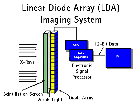
About ten years ago, the linear diode array (LDA) imaging system was introduced into the NDT market. The LDA offered more favorable characteristics in many areas. However, the first LDA systems were very hardware specific and exhibited problems with decreased reliability and increased installation and routine maintenance time. Over the years, improvements were made, and today’s LDA is a technologically advanced imaging system.
This advanced linear diode array imaging system exhibits higher resolution for finer image detail, provides longer life for higher reliability and lower maintenance costs, and uses a 12-bit preprocessor to accommodate a wide dynamic range. Today’s LDA system consists of a PC (standard IBM architecture with Microsoft Windows NT operating system), an SVGA monitor, and a 1,024-element diode array.
The flat panel detector is one of the newer digital technologies available today. Two common types of panels are the amorphous silicon detector (a-Si) and the amorphous selenium detector (a-Se). Amorphous silicon detectors are commonly offered in two pitch sizes:
- 127 micron pitch which is capable of >3.8 LP/mm when measured at the panel.
- 400-micron pitch which is capable of 1.2 LP/mm when measured at the panel.
Flat panels have many of the same inherent characteristics and advantages as the LDA (see figure 1). Unlike the LDA where either the part or the LDA itself need to be in motion to gather data, the flat panel is able to gather information without motion. Flat panel detectors are available in a variety of sizes. The most common sizes range from 4" x 4" to 12" x 16".
| Image Intensifier | Digital Detectors |
| Can generate only 256 grayshades (8-bit data) | Can generate 4096 grayshades (12-bit data) or greater |
| Image contrast reduced due to X-ray scatter | X-ray scatter has negligible effect on image contrast * |
| Blooming at edges results in loss of defect sensitivity | No blooming so edges can be inspected with same resolution as rest of part |
| Increased potential for geometric distortion | Eliminates geometric distortion |
| Relatively slow image acquisition time | Quicker image acquisition time. |
* LDA ONLY.
How the LDA Works
 Groups of 64 diodes are laminated
with a scintillation screen to create X-ray sensitive diodes. X-rays are projected through
the part and received by a single row of diodes. The diodes collect energy for several
milliseconds. The scintillation screen converts the photon energy emitted by the X-ray
tube into visible light on the diodes. The diodes produce a voltage when the light energy
is received. This voltage is amplified, multiplexed, and converted to a 12-bit digital
signal by the interface board and sent to the computer. The diodes are reset and ready to
receive X-rays for the next line of the part to be scanned. Data acquisition
software corrects the incoming data for pixel-to-pixel variations in diode sensitivity.
The data can then either be displayed as an image to an operator or sent to the computer
for analysis.
How the Amorphous Silicon Flat Panel Works
A flat panel detector is a relatively new imaging device. The
panel converts X-ray energy into large-format digital images. A light sensitive photodiode
array (a-Si) is coupled to a scintillation material (GOS screen), which fluoresces during
X-ray bombardment. Upon striking the scintillator material, the X-rays are converted to
visible light, which is detected by the amorphous silicon photodiode array and transformed
into electrical signals. The electrical signals are then extracted from the sensor and a
digital image is produced.
Choices of ADR and How It Works
ADR takes the data provided by the detector, analyzes it, and returns a pass/fail decision.
Golden Standard Extracting Anomalies
This software is based on a Golden Image. The final pass/fail decision depends on the results of a comparison of an acquired production image to a stored Golden Image. A Golden Image database is established by acquiring images of acceptable product.
- A product code which defines a set of scan parameters for a specific product is entered by an operator or determined by an automatic product identification system.
- The system receives and acknowledges the scan parameters then requests and receives an ID to mark the image.
- The system sends the scan parameters to the Panel/LDA and starts the acquisition.
- The Panel/LDA scans the image to the system’s image buffer.
- The acquired image is compared to the image stored in the Golden Image Archive.
- The system requests and receives image results for alignment, feature extract, and feature classification.
- The image results are returned to the operator.
Training a Part
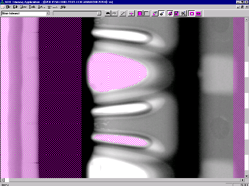
 Training a part usually requires only one revolution of the part. Simple
point and click drawing tools, such as box and paintbrush tools, are used in the training
process. An "undo" function is available. The operator acquires an image and
defines the start and stop locations on the image with a box tool. The operator defines
the regions of non-interest with paintbrush tools. Next, the regions of interest are
defined and the reject level for each defect type is set. Reject levels for each
defect type are indicated with different colors. Defect types available for selection are
cavity, gas, inclusion, and sponge. When this is complete, the trained image or golden
image is saved to the Golden Image database. A golden image need be created only once;
however, if necessary, the operator can easily edit the image in the future.
The Inspection Process
 Proprietary software algorithms capture an image of the part and
align the image to a golden image. The part is compared to the golden image at each region
of interest and for each inspection type and level. Differences are extracted and accept
or reject is based on the extraction results. Data is forwarded to a statistical
database. Flawed parts are identified, and the ADR system signals pass or fail to
the inspection system.
Common defects such as cavity, gas, inclusion, and sponge are detected. Manufacturers are able to define various levels of defects. Defect type and severity are then automatically classified according to these manufacturers-defined criteria. Quantitative measurements of defect size and density provide process feedback.
Neural Network Artificial Intelligence (AI)
The Artificial Intelligence ADR system is designed on the basis of a qualitative image model. The model assumes that the image is defect free and is composed from areas of relative smooth density separated by narrow regions of rapid variations (edges). Edges are not sharp structures in X-ray imagery because of the penetration and divergent rays of the central projection. In the edge regions the noise level is high. The defects are described as local deviations from the smooth intensity given by the continuity of the surface. Gas inclusions or less dense material give rise to local increase in intensity as less X-ray is absorbed. Prior to the development of digital detectors, which have high dynamic range, defects could not be repeatably detected near the edge regions due to high relative noise.
The analysis involved in the identification of defects in complex X-ray imagery take the following steps:
- Edge Detection
- Regions of Interest Network
- Adoptive Reference Subtraction
- Region of Interest Masking
- Defect Identification
- Quality Estimation
The actual location of edges provides information about the orientation of the object, and is used for the trained neural network to estimate the location of the ROI’s that the given geometry contains. Further a segmented edge image is used to mask out the edge regions in order to eliminate false detections in these regions.
The polygonal ROI’s are determined with the edge image as an input; the model is fully emperical and trained by example. Examples are derived by the operator drawing typical configurations in the teach-in phase. The ROI network outputs a mask in which the areas outside the ROI’s are masked out, and so are areas where edge may enter a ROI.
Within each ROI a fixed weight neural network estimates a smooth average intensity surface and subtracts the surface model from the actual noisy surface. The network operates with single sensitivity parameter that is calibrated in the teach mode. The parameter determines the flexibility of the adoptive surface.
The ROI originating from the neural network is used to mask out non-critical areas. Regions of non-interest (RONI) can be specified in ROI’s to minimize false reject sources.
The final imaging process step is to convert the noisy residual resulting from the subtraction in the prior step into a two level image, which is high on the defects and low on the noisy background. A novel non-linear filter based on the so-called Hopfield-Tank neural network is employed to perform the critical task. The algorithm is unique in combining both local smoothing as a low-pass filter and the sensitivity to small defects of a high-pass filter.
The ROI specific configuration of defects are quantified and fed to the final classification system. The classification is based on look-up tables or user standards.
Note: Prior to the development of digital detectors, edges produced unwanted noise due to low dynamic range which is inherent in Image Intensifiers.
Rule Base Using Specific Algorithms
The Rule Base software system is designed to accept/reject from known standards or algorithms. Test programs are loaded either automatically by the test sample recognition module or by the operator using a standard computer and mouse to point and click. The advantage of the automated test sample recognition module is that sorting of different test samples is not required. This allows the system to be fed with all production samples in production, in random order.
Operation sequence:
- Enter or recall a test routine
- Adjust the test position
- Adjust the proper X-ray technique and ADR parameters. Integration, contrast, sensitivity, and image segmentation is adjusted for every X-ray image so that reference defects are recognized true to size and location.
- Enter the maximum allowable defect size and allowable cluster parameters.
- Regular image structures are self-taught by the system by running multiple test samples through the system.
ADR Advantages of a PC Platform over Proprietary Hardware
The open architecture of the PC platform makes many ADR packages affordable, easy to maintain, and easy to upgrade. With Microsoft Windows NT as the operating system and point and click graphical user interface (GUI), the operator interface is user friendly and setup time is lower.
Images can be stored to the system hard drive, CD-ROM, or network drive.
ADR False Accepts and Rejects
 On average, false accepts are < 0.1 percent while false rejects are
< 3 percent. However, normal process variations may sometimes appear to be
defects. It is statistically improbable to find very tiny or marginal flaws and never
reject good product. This is true for human as well as machine inspection.
Conclusion
Choosing the right X-ray source, detector, and automatic defect recognition platform will be dependent upon each manufacturers specific requirement. Automatic defect recognition over operator analysis offers manufacturers a significant competitive edge through labor savings, reduced liability, higher quality product, and improved process controls.
Automatic defect recognition is currently being used in the semi-precious metals, cast aluminum wheel, general-purpose castings, and just recently introduced to the tire industries with excellent results. With continuous improvements in computer processing power, faster inspection times yielding greater throughput is achieved as advanced technology reaches for the new millennium.
[technical_articles_selections_tab.htm]